
Before and After Wisdom Teeth Face Change: Timeline & Evidence
By the Wisdom Teeth Help Editorial Team · Updated August 29, 2026
By the Wisdom Teeth Help Editorial Team · Updated August 29, 2026
Editorial note: This article was checked against the available peer-reviewed facial-volume study and the cited patient-education materials from dental and oral-surgery practices. It has not been independently reviewed by a clinician and does not replace advice from your dentist or oral surgeon.
The short answer: expect temporary puffiness, not a new face shape
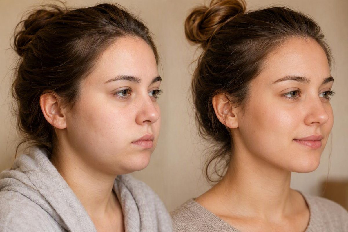
For most people, wisdom-tooth removal is unlikely to produce a lasting, visible change in facial shape. What commonly changes is the face’s temporary appearance during recovery: the cheeks may look wider, rounder, bruised, or uneven because of postoperative inflammation, fluid buildup, and jaw stiffness. These effects generally diminish as healing progresses rather than representing movement of the jaws or cheekbones after wisdom-tooth extraction.
Wisdom-tooth extraction is not supported as a facial-slimming, cheek-hollowing, or jawline-contouring procedure. It removes third molars at the back of the mouth; it does not ordinarily remove facial fat, lift the cheekbones, or reposition the jaws.
It helps to separate three different meanings of “face change”:
- Immediate postoperative change: Swelling, bruising, soreness, fluid retention, and limited jaw movement temporarily alter how the face looks.
- A subjective impression after recovery: Someone may feel that their face looks slimmer, sharper, or more balanced once puffiness or pre-existing inflammation has resolved.
- An objectively measured long-term change: Standardized imaging shows a persistent difference in facial tissue or structure compared with the true preoperative baseline.
Those outcomes are not interchangeable. A dramatic comparison between a swollen day-three photograph and a healed photograph shows that swelling subsided—not that surgery permanently reshaped the face. Similarly, a person can sincerely feel that the jawline looks clearer without having a measurable reduction in facial soft-tissue volume.
The most directly relevant published research is reassuring but limited. In a small 2023 pilot study, researchers detected localized hard-tissue loss on the extraction side but found no statistically significant difference in facial soft-tissue volume between baseline and six months. That result does not prove that no individual can ever perceive a subtle lasting difference; it means the study did not demonstrate a clear, persistent average slimming effect in its sample using three-dimensional facial measurements.
A fair before-and-after assessment therefore requires more than isolated photographs. The timing of swelling, the anatomy involved, the limits of the research, changes in weight or inflammation, and the presence of concerning symptoms all matter.
What your face may look like from surgery day through later healing
There is no universal day on which every patient looks or feels “normal.” Visible appearance, comfort while eating or speaking, jaw movement, and internal socket healing proceed on different schedules. The direction of recovery is generally more useful than a rigid deadline.
Before surgery
Start with your actual baseline. Was one side already puffy because of infection or gum inflammation? Were you holding your jaw differently because of pain? Was mouth opening uncomfortable?
If a pre-existing inflammatory problem resolves after treatment, the healed face may look less swollen than it did before surgery. That is relief of inflammation, not necessarily cosmetic reshaping. If appearance is a significant concern, take neutral baseline photographs before the procedure rather than relying on memory.
First 24 hours
Swelling, soreness, minor oozing, and jaw tightness can begin during this period. The face may not yet be at its puffiest, and one side may look fuller if the teeth or procedures differed between sides. Mild oozing or pink saliva can occur early after extraction.
Follow the operating clinician’s instructions for gauze, medication, food, oral hygiene, and activity. Those directions should take priority over a generic online routine because procedures, health histories, and medications vary.
Days 2–3
Swelling often reaches its maximum approximately 48–72 hours after surgery. The cheeks may look conspicuously round or wide, the jawline can be obscured, and one side may appear larger. Bruising can also become more noticeable after the first day according to oral-surgery recovery guidance.
This is usually the worst stage at which to judge a cosmetic result. A photograph taken near peak swelling is not a valid healed “after” image, nor should it be used as the “before” image in a comparison with a later photograph.
Days 4–7
Swelling and soreness should generally begin trending downward. Bruising may remain visible and can change color as it resolves. Jaw tightness may also persist, temporarily changing how the lower face looks while speaking, smiling, or opening the mouth.
The key word is trend. Improvement does not require every symptom to disappear immediately. A face that remains somewhat puffy but is becoming less swollen is different from swelling that is rapidly enlarging, spreading, or returning after it had started to settle.
Around weeks 1–2
Many people look substantially closer to baseline during this period, although residual puffiness, discoloration, or stiffness can last longer. Recovery varies with the individual and the complexity of the procedure; a practice article authored by an orthodontist describes puffiness as generally resolving within one to two weeks during wisdom-tooth healing.
Two weeks is not a pass-fail deadline. Someone may feel ready for usual activities while still having minor swelling. Conversely, looking normal externally does not mean the extraction sockets have finished healing.
After visible swelling resolves
The gums, sockets, and nearby bone continue repairing internally after the face looks normal. Practice guidance describes internal healing as continuing beyond the initial visible recovery period following wisdom-tooth extraction. That process does not mean the jawline will progressively collapse or the cheeks will become hollow; it means the surgical site is still healing.
When judging appearance, compare the healed face with the genuine preoperative baseline—not with peak swelling and not with a photograph taken while infection or inflammation was distorting one side.
Why removing a back molar usually does not remodel the visible jawline
A wisdom tooth occupies a socket in the tooth-bearing portion of the jaw. After extraction, that localized socket fills and remodels. This is different from cutting, moving, or contouring the larger facial skeleton.
Visible facial shape reflects several overlapping structures:
- The basal maxilla and mandible—the larger upper- and lower-jaw framework
- The cheekbones
- Chewing and facial-expression muscles
- Facial fat compartments, including the cheek fat pads
- Skin and connective tissue
- Overall body composition
Routine wisdom-tooth extraction does not ordinarily reposition the maxilla or mandible. It also does not remove cheek fat. Cosmetic jaw surgery and facial-fat procedures directly alter structures that affect external contour; removing a back molar has a different target and purpose.
Bone does remodel near an extraction socket. The relevant question is not simply whether any hard-tissue change occurs, but whether that change is:
- Large enough to affect the outer contour
- Located where it can influence a visible facial landmark
- Transmitted through muscle, fat, skin, and connective tissue
- Persistent and measurable after healing
A measurable loss of hard-tissue volume close to a socket can therefore coexist with no detectable long-term change in overlying facial soft-tissue volume. This distinction—socket-level remodeling versus face-level contour change—is central to understanding the available evidence.
It also explains why routine extraction is not expected to shift the cheekbones, remove jawline support, or create hollow cheeks. Wisdom teeth are not substitutes for facial fat pads or the broader skeletal framework that supports the cheeks. An oral-surgery practice similarly distinguishes the tooth-bearing extraction area from the parts of the jaw that principally determine visible facial structure when explaining post-extraction anatomy.
The same reasoning applies to fears of facial sagging or accelerated ageing. Those effects have not been established as routine consequences of wisdom-tooth removal. Changes in weight, skin, fat distribution, posture, or natural ageing may occur around the same period, but timing alone does not establish that extraction caused them.
This does not mean local bone remodeling never occurs. It means that local remodeling should not automatically be interpreted as visible loss of cheek or jaw support.
What the six-month 3D study found—and what it cannot prove
The most directly relevant research is a 2023 pilot study measuring facial soft and hard tissues after same-side third-molar extraction. Researchers used structured-light facial scans at baseline, three months, and six months, while cone-beam computed tomography—CBCT—was obtained at baseline and six months.
The study enrolled 24 Chinese adults aged 18–30, with 23 included in the final analysis: 5 men and 18 women. Participants had third molars removed on the same side, and body-weight change was controlled within 2 kilograms. At six months, mean hard-tissue-volume change on the extraction side was approximately −2.33 mL. Extraction-side soft-tissue volume decreased by about 1.396 mL from baseline to three months and then increased by about 1.753 mL from three to six months. Most importantly, there was no statistically significant extraction-side facial soft-tissue-volume difference between baseline and six months, and the researchers detected no correlation between the measured hard- and soft-tissue changes in the peer-reviewed pilot study.
The narrow interpretation is that localized hard-tissue reduction did not produce a clear, persistent average reduction in measured facial soft-tissue volume in this sample. The result does not support wisdom-tooth extraction as a predictable facial-slimming treatment.
The limitations are substantial:
- Small sample: Only 23 participants were analyzed.
- Nonrandomized and nonblinded design: The study was more vulnerable to bias than a large, blinded investigation.
- Predominantly female sample: Eighteen of the 23 analyzed participants were women.
- Narrow age and population: Participants were Chinese adults aged 18–30 who met specific eligibility criteria.
- Specific extraction pattern: The study examined same-side extraction, not every possible combination of one, two, three, or four wisdom teeth.
- Group-level result: The absence of a statistically significant average difference does not prove that no participant perceived a change.
- Limited endpoint: Six-month volume measurements do not answer every question about subtle contours, patient satisfaction, difficult impactions, or different surgical techniques.
It remains a pilot study, however, and the authors called for research with a larger sample. Broader, preferably blinded studies would be needed to estimate how often subtle individual differences occur and whether extraction pattern or surgical complexity matters.
The other clinical sources cited in this article are patient-education materials from dental and oral-surgery practices, not systematic reviews or formal professional guidelines. They provide practical context but should not be treated as equal in evidentiary weight to primary research or individualized clinical advice.
Why before-and-after photos can make the change look dramatic
An ordinary photograph records much more than facial anatomy. It also captures the recovery stage, camera setup, lighting, posture, expression, hairstyle, and the person’s condition that day. Unless those variables are controlled, the image cannot establish structural change.
Common photographic confounders include:
- Camera angle and head tilt
- Camera-to-face distance
- Phone lens or focal length
- Overhead versus front-facing light
- Shadows below the cheekbones or jaw
- Chin position and neck posture
- Smiling, clenching, or relaxing the mouth
- Hairstyle and clothing around the jaw
- Time of day
- Hydration and body-weight differences
None of these effects requires anatomical remodeling.
Timing can create the largest illusion. Compare a photograph taken near peak swelling with one taken several weeks later, and the healed face may look dramatically slimmer. That comparison demonstrates that postoperative puffiness resolved; it does not show that the face became thinner than its true preoperative baseline.
Other non-structural explanations include:
- Resolution of preoperative inflammation: An infected or irritated area may have caused puffiness before surgery.
- Temporary dietary or weight change: Eating differently during recovery may alter facial fullness.
- Reduced muscle guarding: Pain and stiffness can change how someone holds the jaw.
- Uneven healing: One side may swell more when tooth position or surgical difficulty differs.
- Heightened attention: Frequent mirror checks can make minor normal differences seem newly significant.
Dental-practice discussion of postoperative photographs likewise identifies swelling, stiffness, dietary change, weight change, and increased attention to appearance as possible explanations—not proof of skeletal alteration in wisdom-tooth before-and-after images.
For a more useful comparison:
- Use a genuine preoperative photograph as the baseline.
- Take both photographs in the same room and lighting.
- Use the same camera and lens setting.
- Keep the camera at the same distance and height.
- Match head position, chin angle, and posture.
- Use the same neutral facial expression.
- Keep hair away from the cheeks and jaw.
- Compare photographs taken at a similar time of day.
- Account for meaningful body-weight changes.
- Wait until visible swelling has clearly resolved rather than relying on a universal deadline.
Even under controlled conditions, “my face looks more balanced” remains a subjective impression. It is valid as a personal observation, but it is not equivalent to a measured change in three-dimensional facial soft-tissue volume.
Slimmer cheeks, sharper jawline, and other common claims
Myth: Wisdom-teeth removal will slim my face.
Evidence: Extraction is not a predictable slimming procedure. A thinner appearance may follow the resolution of surgical swelling, pre-existing inflammation, muscle tension, or a temporary weight change. The direct pilot study did not find a statistically significant extraction-side soft-tissue-volume difference between baseline and six months.
Myth: Removing the teeth will create a sharper jawline.
Evidence: As puffiness declines, the person’s existing jawline may become easier to see. That is not the same as surgically contouring or repositioning the mandible.
Myth: The teeth support the cheeks, so removal causes hollows.
Evidence: Wisdom teeth do not function as facial fat or as the primary structural support for cheek fullness. Visible cheek contour depends on the broader facial skeleton, muscles, fat, skin, and connective tissue.
Myth: Extraction makes the face sag or age faster.
Evidence: Facial sagging and accelerated ageing are not established routine outcomes of wisdom-tooth removal. Longer-term changes may have other explanations, including weight variation and natural changes in skin or soft tissue. Coincidence in timing does not establish causation.
Myth: Uneven cheeks after surgery mean permanent asymmetry.
Evidence: Early asymmetry commonly reflects unequal swelling. The two sides may differ in tooth position, tissue manipulation, bruising, or inflammatory response. Asymmetry that rapidly worsens, spreads, or occurs with other concerning symptoms should be assessed rather than accepted as a cosmetic outcome.
Myth: Removing all four teeth must change the face more.
Evidence: The direct pilot study assessed same-side extraction. It cannot establish whether removing all four wisdom teeth has a different long-term effect on visible facial contours.
Myth: A difficult impaction or removal of bone definitely changes the face.
Evidence: A more involved procedure may affect short-term swelling and recovery. It would be misleading either to promise such a change or to categorically rule out every subtle individual effect.
Myth: If I see a difference, the measurement must have changed too.
Evidence: Personal perception and objective measurement answer different questions. A person can sincerely believe that the face looks slimmer because inflammation, muscle guarding, weight, posture, or photography changed even when standardized imaging does not show persistent surgery-related soft-tissue loss.
Practice-based patient education also characterizes postoperative puffiness as temporary rather than evidence that facial features have been permanently altered after routine extraction.
Do not remove wisdom teeth for a promised cosmetic result
The available evidence does not support wisdom-tooth extraction solely to slim the cheeks, create hollows, or sharpen the jawline. Surgery may have a clinical purpose when a tooth is causing a specific dental problem, but predictable facial contouring is not an established benefit.
Removal decisions should follow an individualized dental assessment. Findings that may prompt evaluation include:
- Impaction or abnormal eruption
- Recurring pain
- Infection or gum inflammation
- Decay in the wisdom tooth or a nearby tooth
- Cyst development
- Difficulty keeping the area clean
- Possible damage to an adjacent structure
The specific reason matters. Ask the clinician to identify the tooth involved, show what the examination or imaging demonstrates, explain what may happen with observation, and describe the realistic benefit expected from extraction.
Healthy, functional, properly positioned, symptom-free wisdom teeth that can be kept clean may sometimes be monitored rather than automatically removed. Dental imaging can help establish tooth position and inform the recommendation, but it must be interpreted as part of the complete clinical picture. This approach is also described in dental-practice guidance distinguishing problematic teeth from healthy, cleanable teeth that may be observed with periodic assessment.
Useful questions for a dentist or oral surgeon include:
- What specific finding creates a reason to remove this tooth?
- Is there active disease, recurring pain, or documented risk to a neighboring structure?
- Is monitoring reasonable in my case?
- Does the tooth’s position make surgery more complex?
- What are the realistic benefits and risks?
- What temporary swelling or asymmetry should I expect?
- Which symptoms require a same-day call?
- What evidence supports any cosmetic claim being made?
Do not base the decision on a generic assertion that wisdom teeth routinely cause dental crowding, orthodontic relapse, or bite disruption. Any such concern requires a case-specific explanation.
Keep symptom relief separate from cosmetic contouring. If extraction resolves chronic inflammation, the face may look less puffy afterward. That can be a welcome result, but it is not the same as deliberately removing facial volume or reshaping the jaw.
Normal facial swelling versus a reason to call your surgeon
Common early findings include swelling, soreness, bruising, minor oozing, and temporary jaw tightness. These can make the face appear wider or uneven and may limit smiling or mouth opening.
The overall direction matters more than one photograph. Swelling that builds during the first few days and then gradually improves fits the usual postoperative pattern. Swelling that rapidly enlarges, spreads, becomes unusually one-sided, or worsens after starting to settle needs professional attention.
Seek urgent or emergency assessment now if you are having difficulty breathing or swallowing or if swelling is increasing rapidly enough to threaten your airway. Do not wait for a scheduled follow-up or try to judge the problem through photographs.
Contact the treating dentist or oral surgeon promptly for:
- Rapidly worsening or unusually asymmetric swelling
- Fever
- Pus-like drainage
- Severe pain that is worsening rather than easing
- Persistent lip or chin numbness
- Uncontrolled bleeding
Oral-surgery guidance identifies breathing or swallowing difficulty, rapidly increasing swelling, uncontrolled bleeding, fever, drainage, worsening severe pain, and persistent numbness as warning signs requiring urgent professional contact after wisdom-tooth removal.
Dry-socket pain commonly begins several days after extraction and worsens instead of steadily easing. Appearance alone cannot diagnose it. Increasing pain—particularly after initial improvement—deserves a call to the treating clinician.
For ordinary early discomfort, measures such as cold packs during the initial period, head elevation, hydration, and rest may help if they are consistent with the operating clinician’s instructions.
For more context on the typical sequence of recovery, see this wisdom-teeth recovery timeline and dry-socket guide. It is general educational material, not individualized diagnosis or treatment.
Frequently asked questions
Will wisdom-teeth removal make my face permanently thinner?
Probably not. Current direct evidence does not support extraction as a reliable way to reduce facial soft-tissue volume or permanently slim the cheeks. Most early differences reflect swelling, bruising, inflammation, and stiffness. A subtle individual difference cannot be ruled out, but it should not be expected or promised.
When will my face look normal after wisdom-teeth extraction?
Swelling often peaks at approximately 48–72 hours and then begins improving over the following days. Many people look substantially closer to baseline within roughly one to two weeks, but bruising, stiffness, and residual puffiness may last longer. Visible recovery, functional recovery, and internal socket healing do not finish simultaneously according to orthodontic-practice patient guidance.
Can removing all four wisdom teeth change my face more than removing one?
The available direct study cannot answer that question because it examined same-side extraction rather than comparing one-tooth and four-tooth procedures. Removing more teeth may affect the amount and distribution of short-term swelling, but a greater permanent cosmetic change has not been established.
Why does one side of my face look larger after surgery?
The sides may differ in tooth position, extraction difficulty, tissue manipulation, bruising, and inflammatory response. Temporary unevenness can therefore occur. Seek professional assessment if asymmetry is rapidly worsening, unusually severe, spreading, or accompanied by fever, drainage, increasing pain, persistent numbness, uncontrolled bleeding, or difficulty breathing or swallowing.
Can wisdom-teeth removal cause sunken cheeks or facial ageing?
Routine extraction is not supported as an expected cause of sunken cheeks, facial sagging, or accelerated ageing. Wisdom teeth are not facial fat pads and do not ordinarily provide the main support for cheek fullness. Clinic guidance likewise distinguishes temporary postoperative puffiness from permanent loss of facial support after wisdom-tooth removal.
The bottom line is calm and straightforward: the dramatic facial difference most people see shortly after surgery is swelling, not a permanently altered face. Current direct evidence does not support wisdom-tooth removal as a predictable way to slim the cheeks or contour the jaw, although the research is too limited to rule out every subtle individual difference.
Judge appearance only after swelling has resolved, compare photographs under consistent conditions, and choose extraction for individualized clinical reasons rather than cosmetic promises. If swelling, pain, numbness, bleeding, fever, drainage, or breathing or swallowing symptoms are worsening, seek appropriate professional care promptly.