
3rd Molar Extraction: When Is It Needed?
Disease, damage or substantial future risk may favor removal; a painless, disease-free impacted tooth may instead be monitored with planned follow-up.
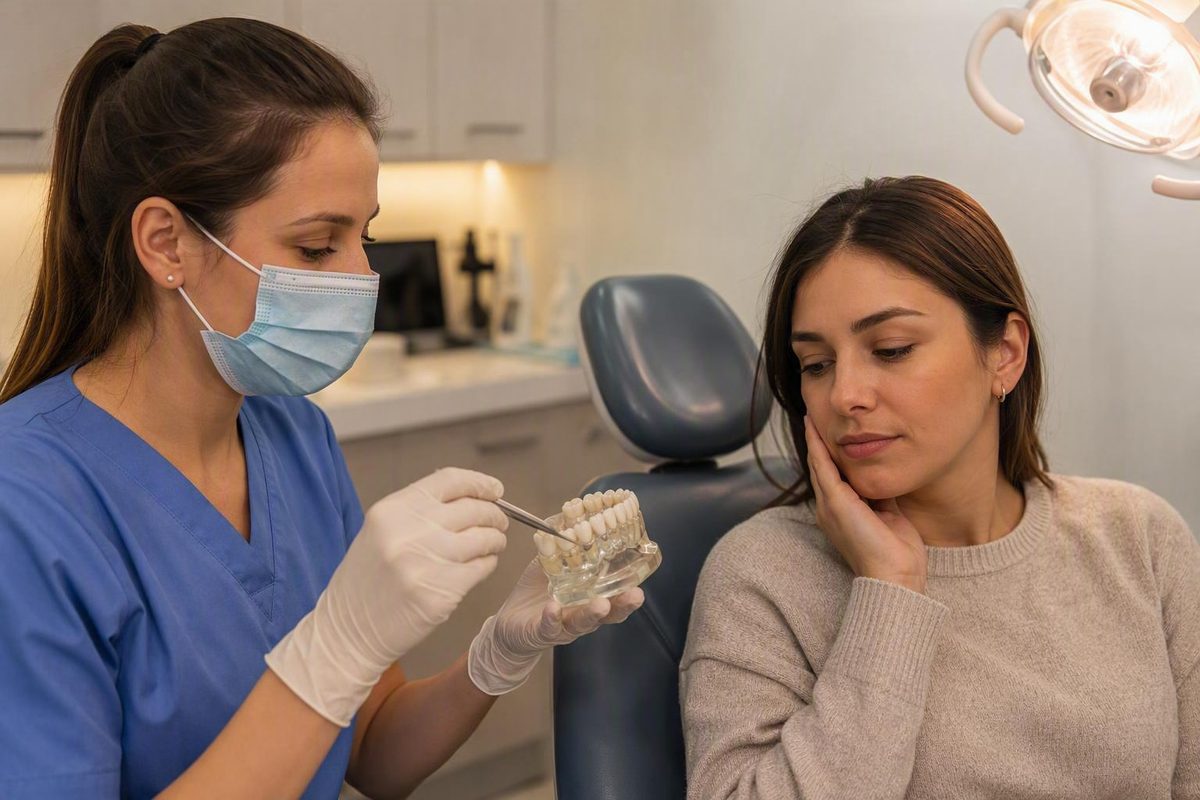
Third molars are wisdom teeth, which usually emerge in the late teens or early twenties. They do not automatically need extraction. Disease, damage, or a substantial future risk often favors treatment, while a painless, disease-free impacted tooth may be suitable for planned monitoring. The right choice depends on examination and imaging findings, the risks of surgery and retention, and whether reliable follow-up is practical.
The short answer: extraction is not automatic
A wisdom tooth may be:
- Erupted: fully through the gum and visible in the mouth.
- Partially erupted: only part of the crown has come through.
- Impacted: unable to emerge normally because of its position, limited space, or an obstruction.
Third molars commonly appear between ages 17 and 25, although they may be visible on dental imaging before eruption. An impacted tooth may remain beneath gum or bone or emerge only partly through the gum, according to the American Association of Oral and Maxillofacial Surgeons’ patient information.
Three separate questions matter:
- Does the tooth cause symptoms?
- Has it erupted?
- Is it diseased or damaging another structure?
An impacted tooth is not necessarily diseased, and a painless tooth is not necessarily healthy.
| Clinical situation | Likely discussion |
|---|---|
| Established disease or damage | Extraction or another treatment is generally favored |
| Elevated future risk without current disease | Individualized comparison of intervention and retention |
| Asymptomatic and disease-free | Active surveillance may be reasonable |
For the narrow question of what to do with an asymptomatic, disease-free impacted wisdom tooth, the evidence does not provide a universal answer. A 2020 Cochrane review found insufficient evidence to determine whether routine removal or retention is superior. It included only two eligible studies, and the relevant evidence was rated low or very low certainty.
Insufficient evidence does not mean the choices are equal, nor does it prove that either approach is universally safer. It means the decision should account for the individual tooth, the patient’s priorities, the feasibility of follow-up, and the uncertainties on both sides.
Findings that may support third-molar extraction
The presence of an impacted tooth is not, by itself, a diagnosis. A professional recommendation should identify a current problem, a meaningful future risk, or a specific treatment-planning need.
Findings that may support extraction include recurrent pericoronitis, tooth decay, periodontal defects, damage to the adjacent second molar, root resorption, cysts, tumors, and other associated disease. A partially erupted tooth can be particularly difficult to clean because gum tissue may cover part of the crown, creating an area where plaque and debris collect and the surrounding tissue becomes inflamed or infected.
The neighboring second molar deserves as much attention as the wisdom tooth. A clinician may look for a deep pocket behind it, bleeding on probing, bone loss, decay, root resorption, or an area that cannot be cleaned effectively. Both retention and extraction can affect the second molar’s periodontal health, so its condition should be documented before choosing a plan. The AAOMS review of third-molar data emphasizes evaluating the third and second molars together, including pocket depth, bleeding, plaque access, and existing bone defects.
| Finding | Why it matters | What the clinician may evaluate next |
|---|---|---|
| Repeated swelling or infection around a partly erupted tooth | May represent recurrent pericoronitis | Gum coverage, drainage, cleaning access, severity, and recurrence |
| Caries in the wisdom tooth | May be difficult to restore or maintain | Depth of decay, restorability, tooth position, and access |
| Decay or a periodontal defect behind the second molar | Retention may complicate cleaning or treatment | Pocketing, bleeding, bone level, plaque access, and second-molar prognosis |
| Root resorption or contact-related damage | May threaten the neighboring tooth | Location and extent of damage on appropriate imaging |
| Cyst, tumor, or other tissue change | Requires diagnosis and management rather than observation alone | Imaging, surgical assessment, and whether tissue examination is needed |
| Obstruction of second-molar eruption | May interfere with a specific orthodontic objective | Tooth position, available space, and the orthodontic plan |
| Relevance to planned jaw surgery | The tooth may affect surgical access or timing | Coordination with the surgical and orthodontic teams |
Pain should prompt evaluation, but pain alone does not establish that a wisdom tooth is responsible. The clinician should identify the source rather than treating the location of discomfort as the diagnosis.
When surveillance may be reasonable
Active surveillance may be considered when a wisdom tooth is asymptomatic, shows no clinical or radiographic disease, is functional or potentially maintainable, and is not judged to present a significant risk to nearby structures.
Surveillance does not mean forgetting about the tooth. It means returning for planned clinical assessment and radiographs selected by the treating clinician. Timing should reflect the tooth’s position, cleaning access, previous findings, medical and dental history, and any changes over time.
Some retained, disease-free third molars may remain problem-free throughout life. Others may erupt, change position, or develop disease later, and that course cannot be predicted confidently for every patient. Possible retention problems include pericoronitis, caries, periodontal deterioration, cystic change, root resorption, and damage to the adjacent second molar.
The comparative evidence remains limited. Very low-certainty evidence suggests an association between retention and greater long-term periodontitis risk at the adjacent second molar, but it does not establish that the retained tooth caused the disease. The studies also failed to measure several important outcomes, including many surgical harms and some possible consequences of retention. The Cochrane review therefore supports shared decision-making and regular assessment when an impacted tooth is retained, not a blanket rule for removal or observation.
Practical feasibility matters. Retention becomes less attractive when a patient is unlikely to attend follow-up, cannot obtain recommended imaging, or cannot keep the area clean. Conversely, a maintainable tooth with stable findings and reliable follow-up may not justify immediate surgery merely because it is impacted.
How a dentist or oral surgeon evaluates the decision
A useful consultation should cover more than whether the tooth “looks stuck.” Expect the clinician to consider:
- Current pain, swelling, bad taste, drainage, or difficulty opening the mouth
- Previous episodes of inflammation or infection
- Medical history, medications, allergies, and relevant dental history
- Whether the tooth is erupted, partly erupted, or fully impacted
- Its angle, depth, root shape, and relationship to nearby structures
- Whether it functions as a useful chewing tooth
- Caries in the third molar or second molar
- Gum pocketing, bleeding, bone support, and plaque access
- Whether the patient can clean and maintain the area
- The health and prognosis of the adjacent second molar
Imaging may help show the crown and roots, associated disease, effects on the second molar, and the relationship of a lower tooth to the inferior alveolar nerve or an upper tooth to the maxillary sinus.
AAOMS recommends combining medical and dental history, examination, imaging, patient-specific removal and retention risks, and discussion with the patient. Its third-molar management statement favors surgery for teeth associated with disease or significant disease risk and active clinical and radiographic surveillance when both are absent. This is professional-association guidance rather than high-certainty comparative research, so it should inform—not replace—individualized decision-making.
Bring these questions to the consultation:
- What exact disease or risk do you see?
- Can you show me the relevant finding on the examination or image?
- What could happen if I monitor the tooth instead?
- What are my anatomy-specific surgical risks?
- Is complete extraction the only reasonable option?
- What follow-up would retention require?
- What change would make you recommend treatment later?
The final plan should combine examination and imaging findings, surgical risk, retention risk, follow-up feasibility, patient preferences, and applicable professional guidance.
Extraction, coronectomy, or surveillance: compare the options
Complete removal is not the only possible management approach. The principal choices are extraction, coronectomy in selected cases, and active surveillance.
| Option | What happens | Potential benefit and limitation | Surgical exposure and follow-up |
|---|---|---|---|
| Complete extraction | The crown and roots are removed | Completely removes the tooth, but does not necessarily resolve every associated periodontal or adjacent-tooth problem | Greatest immediate intervention; postoperative review may be needed |
| Coronectomy | The crown is removed while part of the roots remains | A partial-removal option for selected situations; not appropriate for every diagnosis or anatomy | Still involves surgery; retained roots require an individualized follow-up plan |
| Active surveillance | The tooth remains in place | Avoids immediate surgery, but future disease remains uncertain | No immediate surgical exposure; planned clinical and radiographic follow-up is required |
Complete extraction may be favored when existing disease or damage makes treatment necessary. Its expected benefit should still be compared with anatomy-specific risks, especially if the tooth is close to a nerve, sinus, or compromised second molar.
Coronectomy is a partial-removal option used in selected situations.
AAOMS also lists specialist options such as surgical exposure, tooth repositioning, transplantation, periodontal surgery, and management of associated soft-tissue pathology. These are selected treatment-planning tools, not routine alternatives for every impacted tooth. The association’s management statement presents these approaches as part of a broader individualized management framework.
For each option, ask about the immediate intervention, expected benefit, remaining uncertainty, possible complications, and long-term follow-up burden.
Age, timing, and the crowding question
Extraction may become more technically difficult with age, and postoperative pain, discomfort, or complications may increase. Age therefore changes the risk comparison, but it is not a disease and does not create an automatic indication for extraction.
AAOMS advises making a deliberate decision to remove or continue observing before the middle of the third decade. That is professional guidance, not a requirement to extract every wisdom tooth by a fixed birthday. A retained tooth may remain healthy, while disease that develops later could make subsequent treatment more complex.
The crowding argument should be separated from genuine orthodontic indications. Current comparative evidence does not demonstrate a clinically meaningful benefit from prophylactic third-molar extraction for preventing late lower-incisor crowding or orthodontic relapse. The Cochrane analysis found low-certainty evidence showing no clinically significant effect on dental-arch dimensions. Its limitations mean it cannot prove that the effect is exactly zero, but it does not support crowding prevention as a reliable stand-alone benefit.
That differs from a specific treatment-planning problem, such as a third molar physically obstructing eruption of the second molar or affecting planned orthognathic surgery. If you are receiving orthodontic treatment, ask the orthodontist and surgeon to identify the exact objective and show how the third molar affects it.
Procedure planning and extraction risks
Wisdom-tooth removal is commonly performed as an outpatient procedure with anesthesia. Time and complexity vary with the number of teeth being removed, whether they are erupted or buried, their depth and angle, root anatomy, and their relationship to nerves, the sinus, and adjacent teeth.
The AAOMS patient FAQ notes that an uncomplicated extraction of all four third molars can sometimes take less than an hour. That is a bounded example—not a promise or a typical time for every patient.
Anesthesia arrangements also vary. Obtain procedure-specific instructions directly from the treating office about fasting, medications, an escort, driving, and supervision afterward. Do not assume that instructions for local anesthesia, IV sedation, and general anesthesia are interchangeable.
Potential complications include:
- Pain and swelling
- Bleeding
- Alveolar osteitis, commonly called dry socket
- Infection
- Trismus, or restricted mouth opening
- Gum or bone injury around the second molar
- Temporary or persistent altered sensation
Upper wisdom teeth may be near the maxillary sinus. These relationships can affect the surgical approach, consent discussion, and whether another management option should be considered. A 2015 dental journal editorial describes pain, swelling, bleeding, dry socket, infection, altered sensation, and trismus among reported postoperative problems while emphasizing the need for a specific reason to operate.
Before consenting, ask:
- What is the exact reason for removing this tooth?
- What are the alternatives, including surveillance?
- Which risks are increased by my anatomy or health history?
- What anesthesia is planned?
- How complex do you expect the extraction to be?
- What medication and fasting instructions apply?
- Who should I contact after hours if something changes?
Recovery phases, clot protection, and warning signs
Recovery is better understood in broad phases than as a rigid timetable. Your surgeon’s written instructions take priority because the procedure, closure method, medications, and anesthesia plan vary.
During the first 24 hours: Use steady gauze pressure as instructed, protect the blood clot, and limit activity. Once numbness has resolved, choose cool or soft foods that do not require forceful chewing. Use ice only as directed. Avoid smoking, straws, vigorous spitting, and vigorous rinsing as instructed because suction or force may disturb the clot.
Over the next several days: Some discomfort, swelling, jaw stiffness, and minor bleeding or blood-tinged saliva can occur. Continue the prescribed cleaning, diet, medication, and activity plan. Brushing and rinsing instructions differ between practices, so do not substitute a generic schedule for the one supplied by your surgeon.
During the first week: Symptoms should generally move toward improvement, although day-to-day recovery varies.
Over subsequent weeks: The socket and surrounding tissues continue to heal. Advance food texture, exercise, smoking-related restrictions, and oral hygiene according to the treating team’s instructions rather than a universal calendar.
| Can occur during recovery | Contact the treating team promptly |
|---|---|
| Manageable discomfort | Pain that worsens after initial improvement |
| Swelling that stabilizes and then improves | New or increasing swelling after improvement |
| Minor oozing or blood-tinged saliva | Bleeding that remains uncontrolled despite instructed pressure |
| Temporary jaw stiffness | Fever, pus, or other signs of infection |
| Short-lived numbness immediately after anesthesia | Persistent, worsening, or concerning altered sensation |
For uncontrolled bleeding, fever, pus, worsening pain or swelling after initial improvement, or concerning altered sensation, contact the treating team promptly. Practice-specific post-extraction guidance similarly emphasizes clot protection and directs patients to call for persistent bleeding, increasing symptoms, or prolonged altered sensation.
The surgical office may need to examine the socket.
Wisdom Teeth Help provides attributed general information, not diagnosis, emergency care, or individualized postoperative instructions. Urgent or concerning symptoms require direct contact with the surgical team or emergency services, as appropriate.
What affects the cost of third-molar extraction?
The number of teeth being removed, the degree of impaction, and insurance coverage can affect cost, according to the AAOMS wisdom-teeth FAQ.
Ask for a written estimate and clarify what it includes, such as the consultation, imaging, procedure, anesthesia, pathology if applicable, and postoperative care. Also ask whether the estimate could change after the surgeon reviews the examination and imaging.
Can wisdom teeth grow back after extraction?
No. A third molar that has been completely extracted does not regenerate. In uncommon situations, an additional or supernumerary tooth may exist and later appear to be a “new” wisdom tooth. The same AAOMS patient FAQ notes that preliminary dental imaging can often identify whether extra teeth are present.
The most useful decision prompt is simple: ask the clinician to identify the exact disease or future risk, show you the relevant examination or imaging finding, compare extraction with surveillance or another option, and define the follow-up plan. Disease and damage often make removal reasonable. A painless, disease-free impacted tooth calls for an individualized choice—not an automatic extraction.